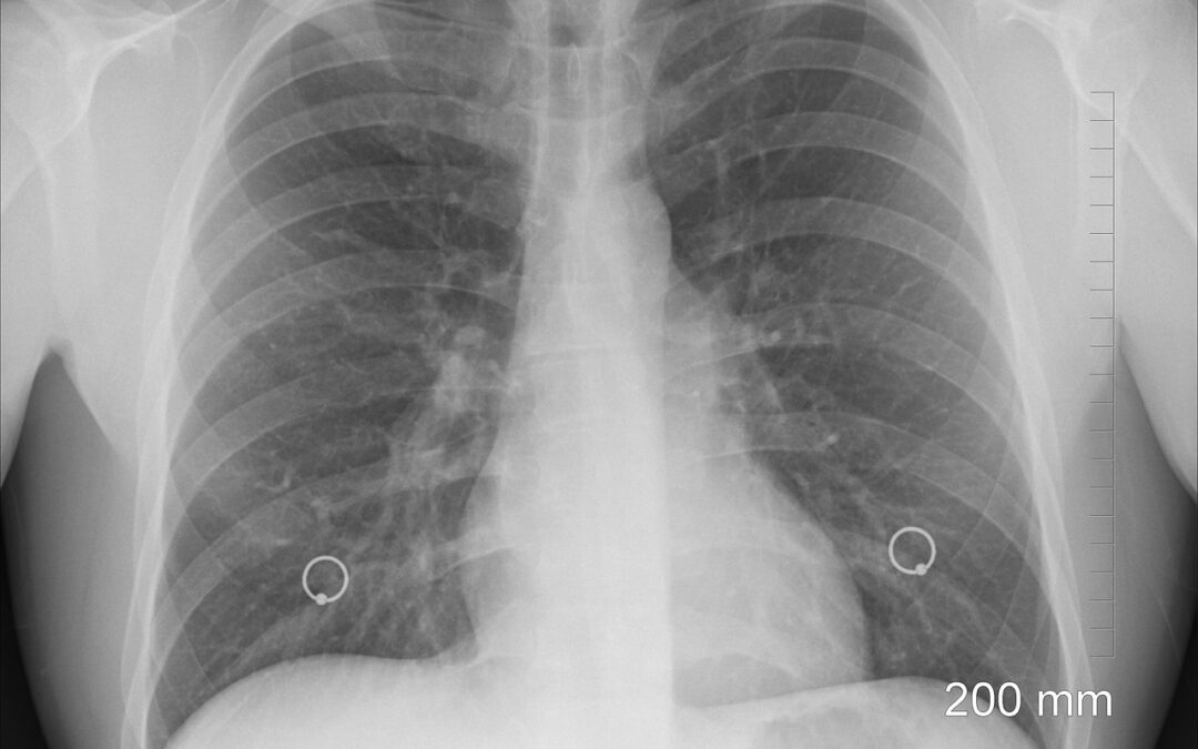
A study published recently in the scientific journal Nature Communications analyzed how a new technology could be used to detect lung cancer at the cellular level. The researchers believe this technology could allow physicians to diagnose lung cancer earlier, opening the door for more effective treatment regimens. Here is a detailed overview of the study and how it could potentially improve outcomes for lung cancer patients.
The Importance of Early Detection
Early detection is key in the treatment of all forms of cancer, as finding cancer early significantly increases the chances of survival. Lung cancer is the leading cause of cancer deaths in the United States, but it can be treated if found early enough. However, the disease becomes nearly impossible to treat after it has spread to other parts of the body.
Treatment most commonly involves a combination of surgery, chemotherapy, and immunotherapy. Cancer researchers have been researching ways to detect lung cancer earlier for decades, as detecting it as early as possible could drastically improve lung cancer outcomes.
Current Challenges in Lung Cancer Screening
Although biopsies are often used as a diagnostic tool for lung and other forms of cancer, this method is severely limited. Lung nodule biopsies have inherently low diagnostic yields, which makes it difficult to differentiate between cancerous and benign nodules.
Currently available medical technology does not allow for real-time diagnostics during biopsies. Instead, oncologists must conduct histopathologic analysis, which can take several days.
Oncologists have responded to these challenges by attempting to develop techniques that could allow cancer to be detected at the cellular level in real time, which could be used to make biopsy-based diagnosis more accurate and improve staging for small nodules.
How Can Technology Detect Lung Cancer at the Cellular Level
The Nature Communications study evaluated a novel method for detecting lung cancer at the cellular level. This new technology could be used to detect the disease at a microscopic level, with much more detail than a standard biopsy and tissue analysis. The researchers used mice models, human tissue samples, and cell cultures to conduct the study.
According to the authors, the study showed potential for high diagnostic accuracy when using a real-time needle-based confocal laser endomicroscopy system and a cancer-targeted molecular imaging agent in tandem. This combination was accurately able to assess malignancy in small lung nodules, which are typically difficult areas for cancer diagnosis.
The researchers believe that this method demonstrated an ability to tell the difference between healthy and cancerous cells at the single-cell level. It also was able to detect cancerous cells in tiny tumors at a width of fewer than 2 centimeters.
Behind the Cancer Detection Technology
The technology evaluated in this study is a combination of a fluorescent dye and tumor-targeted optical tracers. A cancer-targeted near-infrared (NIR) tracer was integrated with a needle-based confocal laser endomicroscopy (nCLE) system that was modified to detect the NIR signal.
The tracer contains an NIR fluorescent dye that specifically targets malignant cells that overexpress a certain type of folate receptor. The combination of the dye and tracer (called NIR-nCLE) was examined as a possible way to detect cancer in real time at the single-cell level during a biopsy.
According to the study, the NIR-nCLE combination resulted in rapid, accurate detection of cancer cells during biopsies with much more diagnostic accuracy than histopathologic analysis.
Limitations of This Study
While the results of this study are encouraging, researchers recognize that the technology is not without its limitations.
Further testing is needed during biopsies to determine how different body and health characteristics could affect the method’s effectiveness. The study specifically mentions respiratory variation, blood circulation, and motion as factors that could influence the efficacy of the technology.
The study also relied on a folate receptor-targeted NIR tracer to detect malignant cells. While it proved effective in detecting lung cancer, this tracer might not be effective in tumors that do not overexpress folate receptor alpha.
Fluorescence intensity signals can be powerful tools for identifying tumors. However, this technology has inherent limitations, such as focal plane position, tissue heterogeneity, receptor expression profile, depth of signal penetration, and the number of labeled cells that are within view.
The study also yielded a small number of false-positive diagnoses in both in vitro and in vivo models. The technology will need to be further developed to prevent these false positives, and further research will need to be conducted to determine what caused them.
Future Research
While the researchers are hopeful that this technology could be used to identify other types of cancer, further research needs to be conducted. Future studies might examine how cancer-targeted agents can be used to improve diagnostic outcomes and patient care. The laboratory plans to work toward determining a fluorescence intensity threshold that can allow for definitive identification of malignancies.
Other cutting-edge technologies could also potentially be used in combination with this treatment method. The researchers posited that machine learning could make image standardization and interpretation possible.

