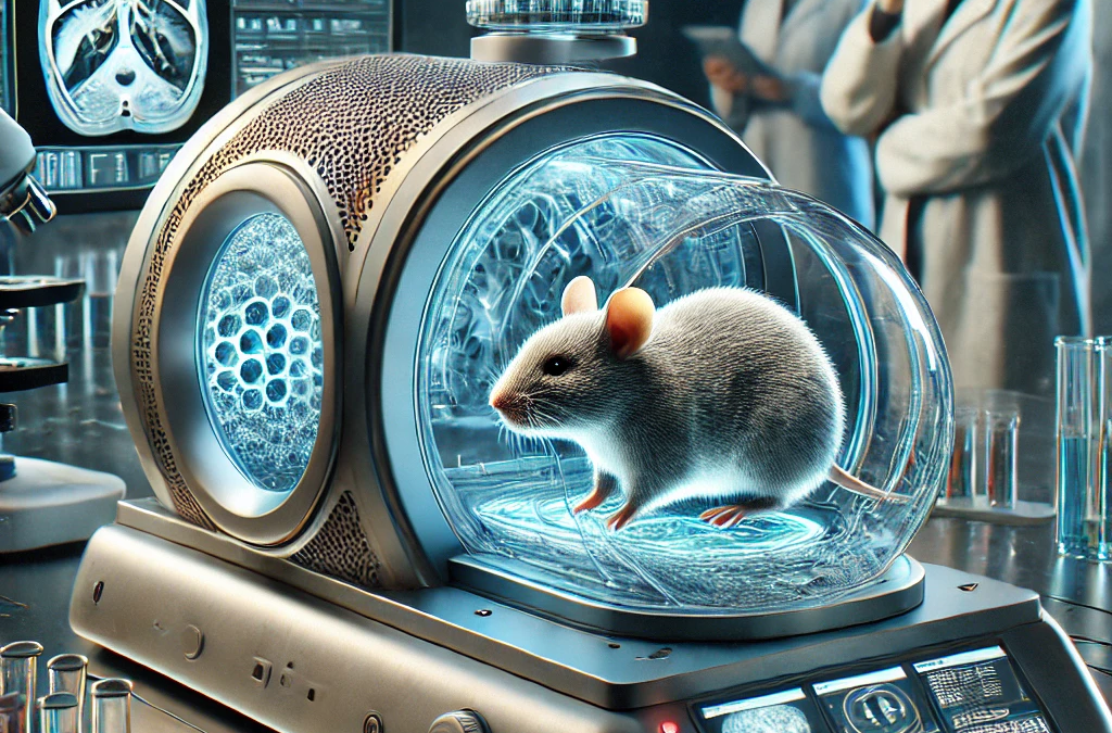
Professor Ali Ertürk and the team at Helmholtz Munich research center have pioneered a new scanning technique involving a transparent mouse that might revolutionize the way cancer drugs are tested. This method illuminates small tumors that remained undetectable by traditional scanning technologies.
The concept of a transparent mouse was first realized by Professor Ertürk in 2018, allowing researchers to observe biological processes with exceptional detail. This most recent development has amplified its potential by using chemicals to highlight specific tissues, resulting in a leap forward in cancer research.
A Game Changer for Drug Testing
The conventional procedure for testing potential cancer drugs involves administering the drug to mice and monitoring tumor progression through traditional scans. After treatment, the mice were scanned again at regular intervals to observe any changes to the size or state of the tumor, thereby estimating the drug’s effectiveness.
However, the newly developed scanning method offers a closer, more detailed look at cancer’s progression or a drug’s efficiency post-mortem. According to Professor Ertürk, this method’s most potent advantage lies in its ability to detect tumors at the single-cell level. This precision is far beyond what MRI and PET scans can achieve.
This pioneering method could potentially improve the efficacy and longevity of cancer patients undergoing treatment by enabling previously invisible minuscule tumors to be addressed. By catching and treating these tiny growths, we might see longer remission periods for human patients, offering a new lease on life to those who previously faced recurring bouts of the disease.
Unique Insights into Cancer Development
Dr. Rupal Mistry from Cancer Research UK explains that this promising scanning technique has the capacity to offer insights into the early stages of cancer and the impact of various treatments. Unlike traditional methods, it enables researchers to visualize tumors within the context of the entire body, offering an unprecedented holistic view of the disease’s progression.
This could prove instrumental in making significant strides in our ability to detect, treat, and prevent cancer. While the technique is currently limited to deceased mice, it is already offering valuable insights into disease development and treatment impacts. It opens a wealth of new information about how the body functions and provides researchers with a more profound understanding of how different drugs affect disease mechanisms.
Broadening the Horizon: Beyond Cancer
Published in journal Nature Biotechnology, this scanning technique’s potential applications extend far beyond cancer research. It holds the promise of enhancing a myriad of medical studies, providing researchers with the extraordinary opportunity to observe biological processes that were previously unseen.
Interestingly, this technique is not restricted to mice. It can be applied to any animal and potentially even to human tissues and organs. While making an entire human body transparent is unlikely in the near future, the potential value of this technique for localized studies in human tissues cannot be overlooked.
The Art and Science of Creating a Transparent Mouse
Creating a transparent mouse is as fascinating as it sounds. It involves removing all fats and pigments from a deceased mouse using a specialized chemical process. The result is a clear, slightly flexible structure that closely resembles a plastic toy, with all organs and nerves intact but nearly invisible.
To further refine this method, Professor Ertürk’s team introduced antibodies into the process. These molecules are known to bind to specific tissues, thereby highlighting areas of interest for study under the microscope. This enhancement enabled the team to generate comprehensive 3D images of various body systems, including the nervous, digestive, and lymphatic systems.
Implications for the Future of Medical Research
This innovative scanning technique presents several significant advantages over existing methods. Firstly, it allows researchers to study diseases in the context of the whole body, improving our understanding of the effects of different drugs and treatments on a comprehensive scale.
The 3D images generated by this method are stored online, creating a virtual library that can be accessed by researchers worldwide. This drastically reduces the need for repeated experiments and potentially minimizes the use of lab animals by a factor of 10, making it not only a scientific breakthrough but also an ethical one.
Dr. Nana-Jane Chipampe from the Wellcome Sanger Institute expressed enthusiasm about this technology’s potential to revolutionize our understanding of human cell development. This scanning technique could enable researchers to observe intricate details in 3D, which is a significant upgrade from studying thin slices of tissue under a microscope.
Professor Muzlifah Haniffa, who is leading a project to map every cell in the human body online, believes that this new scanning technique could be an invaluable tool across various fields of medical research. She envisions a future where this groundbreaking technology accelerates the pace of medical research, propelling us into a new era of scientific discovery and understanding.